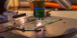

Histopathology, the study of tissues affected by disease, can be very useful in making a diagnosis and in determining the severity and progression of a disease. Understanding the normal structure and function of different tissues is essential for interpreting the changes that occur during disease. This course introduces the basic principles that apply to the preparation of microscope sections. It also shows how to identify a number of human tissues and interpret the changes that occur in disease.
Learning outcomes
After studying this course, you should be able to:
- define all the terms given in bold
- outline key features of a number of pathological processes
- relate the histological appearance of affected tissues to the underlying pathology
- recognise the histological appearance of a number of pathological tissues
- understand how sections can be photographed, presented and reported.
Course Curriculum
| 1. Pathological processes - inflammation and infection | |||
| Pathological processes – inflammation and infection | 01:30:00 | ||
| Acute inflammation | 01:30:00 | ||
| Autoimmunity | 01:30:00 | ||
| Hypersensitivity | 00:30:00 | ||
| Scarring and fibrosis | 01:30:00 | ||
| Wound healing, angiogenesis and tissue regeneration | 01:30:00 | ||
| 2. Pathological processes - neoplasia | |||
| Hyperplasia, dysplasia and neoplasia | 01:30:00 | ||
| Metastasis | 01:30:00 | ||
| 3. Pathological processes - cell death | |||
| Apoptosis and necrosis | 01:30:00 | ||
| Thrombosis and embolism | 01:30:00 | ||
| Degenerative diseases and storage disorders | 01:30:00 | ||
| 4. Photography and reporting | |||
| Image acquisition | 01:30:00 | ||
| Acknowledgements – Histopathology | 00:20:00 | ||
Related Courses

Neuroscience 101
FREE
Brain Dissection
FREE
3 STUDENTS ENROLLED
